سرطان الغدة النخامية
| Pituicytoma | |
|---|---|
 | |
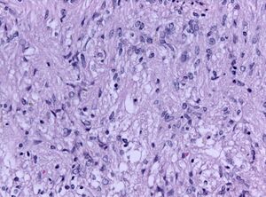
| Biopsy specimen of a pituicytoma of the posterior pituitary gland (H&E stain, x200 magnification) | |
| التخصص | علم الأورام |
ورم الغدة الدرقية هو ورم نادر في المخ. ينمو في قاعدة الدماغ من الغدة النخامية . يُعتقد أن هذا الورم مشتق من الخلايا المتنيّة للفص الخلفي للغدة النخامية ، والتي تُسمى الخلية النخامية. يعتقد بعض الباحثين [1]أنها تنشأ من خلايا الجريب في الفص الأمامي من الغدة النخامية. على هذا النحو ، هو ورم دبقي منخفض الدرجة. يحدث عند البالغين وتشمل الأعراض اضطرابًا بصريًا وخللًا في وظائف الغدد الصماء . غالبًا ما يتم الخلط بينهم وبين أورام الغدة النخامية التي لها عرض مماثل وتحدث في نفس الموقع. يتكون العلاج من الاستئصال الجراحي ، وهو علاج شفائيفي معظم الحالات.[2]
المصادر
- ^ Cenacchi, G.; Giovenali, P.; Castrioto, C.; Giangaspero, F. (July 2001). "Pituicytoma: ultrastructural evidence of a possible origin from folliculo-stellate cells of the adenohypophysis". Ultrastructural Pathology. 25 (4): 309–312. doi:10.1080/019131201753136331. ISSN 0191-3123. PMID 11577776.
- ^ Feng M, Carmichael JD, Bonert V, Bannykh S, Mamelak AN (2014). "Surgical management of pituicytomas: case series and comprehensive literature review". Pituitary. 17 (5): 399–413. doi:10.1007/s11102-013-0515-z. PMID 24037647.
المراجع
- Brat D, Scheithauer B, Staugaitis S, Holtzman N, Morgello S, Burger P (2000). "Pituicytoma: A distinctive low grade glioma of the neurohypophysis". Am J Surg Pathol. 24: 362–368. doi:10.1097/00000478-200003000-00004.
- Cenacchi G, Giovenali P, Castrioto C, Giangaspero F (2001). "Pituicytoma: Ultrastructural evidence of a possible origin from folliculo-stellate cells of the adenohypophysis". Ultrastruct Pathol. 25 (4): 309–312. doi:10.1080/019131201753136331. PMID 11577776.
- Secci F, Merciadri P, Criminelli Rossi D, D'Andrea A, Zona G (2012). "Pituicytomas: radiological findings, clinical behaviour and surgical management". Acta Neurochirurgica. 154 (4): 649–657. doi:10.1007/s00701-011-1235-7.
- Danila DC, Zhang X, Zhou Y, Dickerson GR, Fletcher JA, Hedley-Whyte ET, Selig MK, Johnson SR, Klibanski A (2000). "A human pituitary tumor-derived folliculo-stellate cell line". J Clin Endocrinol Metab. 85 (3): 1180–1187. doi:10.1210/jc.85.3.1180.
- Figarella-Branger D, Dufour H, Fernandez C, Bouvier-Labit C, Grisoli F, Pellissier JF (2002). "Pituicytomas, a mis-diagnosed benign tumor of the neurohypophysis: Report of three cases". Acta Neuropathol. 104: 313–319. doi:10.1007/s00401-002-0557-1.
- Hurley T, D'Angelo C, Clasen R, Wilkinson S, Passavoy R (1994). "Magnetic resonance imaging and pathological analysis of a pituicytoma: Case report". Neurosurgery. 35: 314–317. doi:10.1097/00006123-199408000-00021.
- Inoue K, Couch EF, Takano K, Ogawa S (1999). "The structure and function of folliculo-stellate cells in the anterior pituitary gland". Arch Histol Cytol. 62 (3): 205–218. doi:10.1679/aohc.62.205. PMID 10495875.
وصلات خارجية
| Classification | |
|---|---|
| External resources |
This article contains content from Wikimedia licensed under CC BY-SA 4.0. Please comply with the license terms.