خلية قاعدية
| Basophil | |
|---|---|
.png&filetimestamp=20200330131153&) 3D rendering of a basophil | |
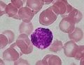
 Basophil among red blood cells | |
| Details | |
| النُطق | /ˈbeɪsəfɪl, -z-, -oʊ-/[1][2] |
| الجهاز | Immune system |
| المُعرفات | |
| MeSH | D001491 |
| TH | TH {{{2}}}.html HH2.00.04.1.02022 .{{{2}}}.{{{3}}} |
| FMA | 62862 |
| المصطلحات التشريحية | |
الخلية القاعدية أو كريات الدم البيضاء القاعدية إنگليزية: Basophil أحد أنواع الخلايا البيضاء وأقلهم عدداً، إذ تمثل نحو 0.5% إلى 1% من خلايا الدم البيضاء في الجهاز الدوري.[3] However, they are the largest type of granulocyte. They are responsible for inflammatory reactions during immune response, as well as in the formation of acute and chronic allergic diseases, including anaphylaxis, asthma, atopic dermatitis and hay fever.[4] They also produce compounds that co-ordinate immune responses, including histamine and serotonin that induce inflammation, heparin that prevents blood clotting,[5] although there are less than that found in mast cell granules.[6] It used to be thought that basophils that have migrated from blood into their resident tissues (connective tissue) are known as mast cells, but this is no longer thought to be the case.[7]
Basophils were discovered in 1879 by German physician Paul Ehrlich, who one year earlier had found a cell type present in tissues that he termed mastzellen (now mast cells).[8] Ehrlich received the 1908 Nobel Prize in Physiology or Medicine for his discoveries.
The name comes from the fact that these leukocytes are basophilic, i.e., they are susceptible to staining by basic dyes, as shown in the picture.
البنية
Basophils contain large cytoplasmic granules which obscure the cell nucleus under the microscope when stained. However, when unstained, the nucleus is visible and it usually has two lobes.[9] The mast cell, another granulocyte, is similar in appearance and function. Both cell types store histamine, a chemical that is secreted by the cells when stimulated. However, they arise from different branches of hematopoiesis, and mast cells usually do not circulate in the blood stream, but instead are located in connective tissue. Like all circulating granulocytes, basophils can be recruited out of the blood into a tissue when needed.
الوظيفة

Basophils appear in many specific kinds of inflammatory reactions, particularly those that cause allergic symptoms. Basophils contain anticoagulant heparin[citation needed], which prevents blood from clotting too quickly. They also contain the vasodilator histamine, which promotes blood flow to tissues. They can be found in unusually high numbers at sites of ectoparasite infection, e.g., ticks. Like eosinophils, basophils play a role in both parasitic infections and allergies.[10] They are found in tissues where allergic reactions are occurring and probably contribute to the severity of these reactions. Basophils have protein receptors on their cell surface that bind IgE, an immunoglobulin involved in macroparasite defense and allergy. It is the bound IgE antibody that confers a selective response of these cells to environmental substances, for example, pollen proteins or helminth antigens. Recent studies in mice suggest that basophils may also regulate the behavior of T cells and mediate the magnitude of the secondary immune response.[11]
صور اضافية
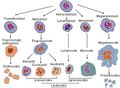
Blood cell lineage
بقعة بديلة من خلية قاعدية


انظر أيضاً
- حساسية
- Diamine oxidase
- Eosinophil
- Food intolerance
- هيستامين
- Histamine intolerance
- Histamine N-methyltransferase or HNMT
- Mast cell
المراجع
- ^ قالب:MerriamWebsterDictionary
- ^ قالب:OxfordDictionaries.com
- ^ "Blood differential test". Medline Plus. U.S. National Library of Medicine. Archived from the original on 21 April 2016. Retrieved 22 April 2016.
- ^ Mukai K, Galli SJ (2013). Basophils. Vol. Online. doi:10.1002/9780470015902.a0001120.pub3. ISBN 978-0470016176. Archived from the original on 2016-05-01.
{{cite book}}:|journal=ignored (help) - ^ Khurana (2009). Textbook Of Medical Physiology (2nd ed.). Elsevier. p. 180. ISBN 978-81-8147-850-4. Archived from the original on 2018-05-04.
- ^ Stone KD, Prussin C, Metcalfe DD (February 2010). "IgE, mast cells, basophils, and eosinophils". The Journal of Allergy and Clinical Immunology. 125 (2 Suppl 2): S73-80. doi:10.1016/j.jaci.2009.11.017. PMC 2847274. PMID 20176269.
- ^ Franco CB, Chen CC, Drukker M, Weissman IL, Galli SJ (April 2010). "Distinguishing mast cell and granulocyte differentiation at the single-cell level". Cell Stem Cell. 6 (4): 361–8. doi:10.1016/j.stem.2010.02.013. PMC 2852254. PMID 20362540.
- ^ Blank U, Falcone FH, Nilsson G (September 2013). "The history of mast cell and basophil research - some lessons learnt from the last century". Allergy. 68 (9): 1093–101. doi:10.1111/all.12197. PMID 23991682.
- ^ "Basophil". medcell.med.yale.edu.
- ^ Voehringer D (December 2009). "The role of basophils in helminth infection". Trends in Parasitology. 25 (12): 551–6. doi:10.1016/j.pt.2009.09.004. PMID 19782643.
- ^ Nakanishi K (December 2010). "Basophils as APC in Th2 response in allergic inflammation and parasite infection". Current Opinion in Immunology. 22 (6): 814–20. doi:10.1016/j.coi.2010.10.018. PMID 21095110.