تخطيط الصدى
| تخطيط الصدى Medical ultrasonography | |
|---|---|
| تدخل | |
| ICD-10-PCS | B?4 |
| ICD-9-CM | 88.7 |
| MeSH | D014463 |
| OPS-301 code: | 3-03...3-05 |
تخطيط الصدى Diagnostic sonography (ultrasonography) أو الموجات فوق الصوتية أو موجات دوبلر التصواتية أو الموجات فوق السمعية هي صور تستخدم الموجات فوق الصوتية وهي أعلى من حدود القدرة السمعية للإنسان التي تبدأ من 20,000 هرتز، أي أنها موجات ذات ترددات عالية نسبيا بحيث أن الأذن البشرية لا تسطيع أن تسمعها.
الاسم الدارج للموجات التصواتية (فوق الصوتية)هو الموجات الصوتية مع أنه لا يمثل حقيقة الموجات المستخدمة. وتستخدم في تشخيص والعلاج أيضا فهي تقوم بتصوير الأنسجة والأعضاء وغيرها ويشيع استخدامها في طب النساء والتوليد.
تطبيقات التشخيص


- متابعة وتصوير الجنين أثناء فترة الحمل.
- تشخيص عدد من أمراض القلب والصمامات القلبية، ومن فوائد هذه الطريقة أن التصوير يتم أثناء عمل القلب، وتكون الصور وظيفية، أي أثناء أداء القلب والصمامات لوظائفها الحيوية.
- تشخيص عدد من الأمراض البطنية، مثل حصى المرارة، التهابات المرارة، حصى الكلى، التهابات الأمعاء، الكشف عن التغيرات المرضية في الكبد والأمعاء والبنكرياس... الخ من الأمراض المتعددة التي تصيب البطن.
- الكشف المبكر عن حالات خلع الورك عند المواليد الجدد.
استخدامات الموجات التصواتية (فوق الصوتية) العلاجية

من الاستخدامات المهمة للموجات التصواتية هو استخدامها في الطب وبالذات في الكشف عن الجنين في بطن أمه وهل هو حي أم ميت، حيث أنه عندما يسلط الطبيب مصدر تلك الموجات على رحم الام تنعكس تلك الموجات وعندما يكون قلب الجنين ينبض فان زمن انعكاس أو ارتداد تلك الموجات يختلف تبعا لانقباض عضلة قلب الجنين وانبساطها وذلك يعود لتغيير بسيط في المسافة التي تقطعها الموجة قبل أن ترتد، مما يعطي الجهاز المستقبل للموجات المنعكسة الفرصة لتسجيل تلك النبضات وبالتالي تظهر على شاشة الطبيب حركة القلب أي أن الجنين يكون حيا، لكن لو كان الجنين ميتا فإن الموجات ترتد في نفس الزمن.كما ان تستخدم الموجات فوق الصوتية في تشخيص اورام البطن والحوض وفي الكشف عن العديد من امراض الجهاز الهضمي والبولي وكذا في فحص الغدة الدرقية والثدى إلى جانب فحص الخصية ويستخدم نظام خاص وهو الدوبلير في معاينة جريان الدم في الاوعية الدموية لذا فهو يستخدم في تشخيص امراض الاوعية الدموية.[3]
وللموجات فوق السمعية استخدامات أخرى عديدة حيث تستخدم في تقدير عمق البحار والمحيطات والكشف عن التجمعات السمكية والجبال الجليدية في البحار والمحيطات ويستخدم القانون التالي في تقدير عمق البحار والمحيطات : المسافة=(سرعة موجة الصوت ×الزمن)÷2
وتستخدم أيضا هذه الموجات في فحص لحام المعادن والمسبوكات حيث يتم تسليط هذه الموجات باستخدام أجهزة خاصة وقياس شدة الموجات المنعكسة وبالتالي يمكن الكشف عن المناطق التي لم يكتمل لحامها جيدا أو التي تحتوي فقاعات من الهواء.
كما تستخدم الموجات فوق السمعية في تعقيم المواد الغذائية وتستخدم في المجالات الطبية في تفتيت حصيات الكلى والحالبين.
يستخدم التخطيط بالصدى لدراسة الحالات الطبية المختلفة:
| الحالة | الوصف | انظر أيضاً |
|---|---|---|
| التخدير | Ultrasound is commonly used by anesthesiologists (Anaesthetists) to guide injecting needles when placing local anaesthetic solutions near nerves | |
| طب القلب | تخطيط صدى القلب، هو فحص القلب باستخدام تقنية الموجات فوق الصوتية. | انظر تخطيط صدى القلب |
| طب الطوارئ | Point of care ultrasound has many applications in the Emergency Department, including the Focused Assessment with Sonography for Trauma (FAST) exam for assessing significant hemoperitoneum or pericardial tamponade after trauma. Ultrasound is routinely used in the Emergency Department to expedite the care of patients with right upper quadrant abdominal pain who may have gallstones or cholecystitis. | انظر FAST exam |
| طب الجهاز الهضمي | In abdominal sonography, the solid organs of the abdomen such as the pancreas, aorta, inferior vena cava, liver, gall bladder, bile ducts, kidneys, and spleen are imaged. Sound waves are blocked by gas in the bowel and attenuated in different degree by fat, therefore there are limited diagnostic capabilities in this area. The appendix can sometimes be seen when inflamed (as in e.g.: appendicitis). | |
| طب النساء والولادة | انظر gynecologic ultrasonography | |
| تقنية نانوية | for basic assessment of intracerebral structural abnormalities, bleeds, ventriculomegaly or hydrocephalus and anoxic insults (Periventricular leukomalacia). The ultrasound can be performed through the soft spots in the skull of a newborn infant (Fontanelle) until these completely close at about 1 year of age and form a virtually impenetrable acoustic barrier for the ultrasound. The most common site for cranial ultrasound is the anterior fontanelle. The smaller the fontanelle, the poorer the quality of the picture. | Intracerebral: see Transcranial Doppler |
| طب الجهاز العصبي | for assessing blood flow and stenoses in the carotid arteries (Carotid ultrasonography) and the big intracerebral arteries | see Carotid ultrasonography. Intracerebral: see Transcranial Doppler |
| طب التوليد | Obstetrical sonography is commonly used during pregnancy to check on the development of the fetus. | انظر obstetric ultrasonography |
| طب العيوم | انظر A-scan ultrasonography, B-scan ultrasonography | |
| طب الجهاز البولي | to determine, for example, the amount of fluid retained in a patient's bladder. In a pelvic sonogram, organs of the pelvic region are imaged. This includes the uterus and ovaries or urinary bladder. Males are sometimes given a pelvic sonogram to check on the health of their bladder, the prostate, or their testicles (for example to distinguish epididymitis from testicular torsion). In young males, it is used to distinguish more benign testicular masses (varicocele or hydrocele) from testicular cancer, which is still very highly curable but which must be treated to preserve health and fertility. There are two methods of performing a pelvic sonography – externally or internally. The internal pelvic sonogram is performed either transvaginally (in a woman) or transrectally (in a man). Sonographic imaging of the pelvic floor can produce important diagnostic information regarding the precise relationship of abnormal structures with other pelvic organs and it represents a useful hint to treat patients with symptoms related to pelvic prolapse, double incontinence and obstructed defecation. It is used to diagnose and, at higher frequencies, to treat (break up) kidney stones or kidney crystals (nephrolithiasis).[4] | |
| Musculoskeletal | tendons, muscles, nerves, ligaments, soft tissue masses, and bone surfaces | |
| الجهاز القلبي الوعائي | To assess patency and possible obstruction of arteries Arterial sonography, diagnose DVT (Thrombosonography) and determine extent and severity of venous insufficiency (venosonography) | Intravascular ultrasound |
التطبيقات العلاجية
من الصوت للصورة
انتاج موجة صوتية

تلقي الأصداء
تكوين الصورة
إظهار الصورة
الصوت في الجسم

طرق التصوير الصوتي
توسعات
التخطيط الصدى الدوپلري


تكون الصورة عادةً ملونة، فيظهر الدم اما بلون أحمر أوأزرق حسب اتجاه الجريان بالنسبة للمجس الفاحص. ويقدم الجهاز معلومات ممتازة في تقييم صمامات القلب وارتفاع الضغوط الدموية في الأوعية الدموية.
Contrast media
تخطيط الصدى الانضغاطي
نقد
المميزات
العيوب
المخاطر والآثار الجانبية
دراسات الأمان على التخطيط بالصدى
الانتشار
التاريخ
انظر أيضاً
- Emergency ultrasound
- 3D ultrasound
- Duplex ultrasonography
- Doppler fetal monitor
- Global Library of Women's Medicine
- Polybiography
- Prenatal nutrition and birth weight
- Handedness - alleged relation to ultrasound
معرض الصور
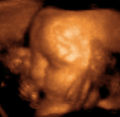
صورة ثلاثية الأبعاد لجنين عمره 29 أسبوع.
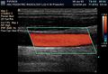
صورة لمقطع عرض شاشة.
تحليل لسرعة جريان الدم بالأوعية.

المصادر
- ^ Dubose, T. J. (1985). "Foetal Biometry: Vertical Calvarial Diameter and Calvarial Volume". Journal of Diagnostic Medical Sonography. 1 (5): 205. doi:10.1177/875647938500100504.
- ^ "3D BPD Correction". July 2000. Retrieved 2008-09-27.
- ^ نبذة تاريخية عن الموجات فوق الصوتية. عن الموقع الإلكتروني للدكتور نجيب ليوس. (بحسب عرض 27.6.2009).
- ^ Sonography of the female pelvic floor Clinical indications and techniques
وصلات خارجية
- American Institute of Ultrasound in Medicine Professional Association
- About the discovery of medical ultrasonography
- History of medical sonography (ultrasound)
- Procedures in Ultrasound (Sonography) for patients, from RadiologyInfo.org
- The Global Library of Women's Medicine Imaging in Obstetrics and Gynecology Link. Non-profit offering freely downloadable expert material for healthcare professionals.
- Careers in the vascular ultrasound field
- Sonography of the female pelvic floor:clinical indications and techniques Illustrate the clinical utility of this non-invasive diagnostic technique.
- How to Become an Ultrasound Technician – a wiki article on becoming an ultrasound technician.