ملف:Hypacrosaurus & emu chondrocytes.jpeg

حجم هذه المعاينة: 800 × 349 بكسل. البعد الآخر: 1٬654 × 722 بكسل.
الملف الأصلي (1٬654 × 722 بكسل حجم الملف: 189 كيلوبايت، نوع MIME: image/jpeg)
وصف قصير
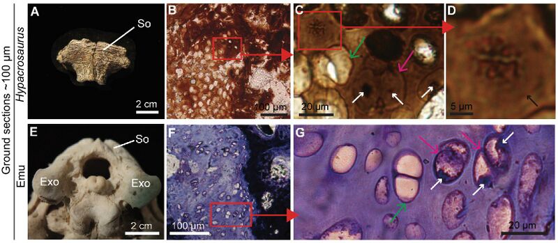
| Description | Ground section of Hypacrosaurus (MOR 548) supraoccipital shows exceptional histological preservation of calcified cartilage. (A) An isolated supraoccipital (So) of Hypacrosaurus in dorsal view. (B–D) Ground section of another So showing calcified cartilage with hypertrophic chondrocyte lacunae. (C) Some cell doublets appear empty (green arrow), but others (pink arrow) present darker, condensed material consistent in shape and location with a nucleus (white arrows). (D) Dark, condensed, and elongated material with morphological characteristics of metaphase chromosomes. The limit of the cell lacuna is visible (black arrow). (E) Caudal view of a juvenile emu skull (∼8–10 months old) showing the So and exoccipitals (Exo) in articulation. (F, G) Ground section (stained with Toluidine blue) of calcified cartilage from this emu skull showing cell doublets (pink arrows) with remnants of nuclei (white arrows) and others without intracellular content (green arrow). |
| Date | 2020-01-12 |
| Source | Bailleul, A. M.; Zheng, W.; Horner, J. R.; Hall, B. K.; Holliday, C. M.; Schweitzer, M. H. (2020). "Evidence of proteins, chromosomes and chemical markers of DNA in exceptionally preserved dinosaur cartilage". National Science Review. 7 (4): 815−822. doi:10.1093/nsr/nwz206. |
| Author | Alida M Bailleul, Wenxia Zheng, John R Horner, Brian K Hall, Casey M Holliday & Mary H Schweitzer |
ترخيص
تاريخ الملف
اضغط على زمن/تاريخ لرؤية الملف كما بدا في هذا الزمن.
| زمن/تاريخ | صورة مصغرة | الأبعاد | مستخدم | تعليق | |
|---|---|---|---|---|---|
| حالي | ★ مراجعة معتمدة 23:51، 28 نوفمبر 2023 | | 1٬654 × 722 (189 كيلوبايت) | Pastakhov (نقاش | مساهمات) | Upload https://upload.wikimedia.org/wikipedia/commons/a/ab/Hypacrosaurus_%26_emu_chondrocytes.jpeg |
لا يمكنك استبدال هذا الملف.
وصلات
لا يوجد صفحات تصل لهذه الصورة.