مجهرية التموقع المنشط ضوئياً
مجهرية التموقع المنشط ضوئياً Photo-activated localization microscopy (PALM أو fPALM)[1][2] and stochastic optical reconstruction microscopy (STORM)[3] هي حقل عريض (كمقابل لتقنيات مسح النقطة مثل مجهرية المسح بالليزر متحد البؤر)، من وسائل تصوير المجهرية الفلورية تسمح بالحصول على صور بدقة تفوق حد الحيود. طرحت الوسائل عام 2006 في أعقاب ظهور الوسائل المجهرية البصرية فائقة الدقة، وأختيرت كوسائل العام في 2008 على مجلة نيتشر ميثودز.[4]
تطوير مجهرية التموقع المنشط ضوئياً كوسيلة تصوير فيزيائية حيوية مستهدفة عززها بشكل كبير اكتشاف أنواع جديدة وهندسة طفرات الپروتينات الفلورية التي تعرض الصباغة الضوئية القابلة للمراقبة، مثل الپروتينات الفلورية الخضراء المنشطة ضوئياً. ومع ذلك، فالتطوير المصاحب لمجهرية التموقع المنشط ضوئياً، يتشارك نفس المبدأ الأساسي المستخدمة في أصباغ السيانين المقترنة.
جزيء واحد من الزوج (يسمى المنشط)، عند تنشيطه للوصول إلى الحد الأقصى من امتصاصه، يعمل على تنشيط جزئيات أخرى (تسمى reporter) إلى الحالة الفلورية.
العدد المتنامي من الأصباغ يستخدم في PALM, STORM والتقنيات المتعلقة، كلاً من الفلورفلورات العضوية والپروتينات الفلورية. بعضها تكون متوافقة مع تصوير الخلية الحية، others allow faster acquisition or denser labeling. اختيار فلوروفور معين يعتمد في النهاية على التطبيق وعلى خصائصه الفيزيائية الضوئية الكامنة.[5]
خضعت كل من هذه التقنيات لتطورات تقنية كبيرة،[6] خاصة فيما يتعلق بالسماح بالتصوير متعدد الألوان والتوسع إلى الأبعاد الثلاثة، مع أفضل دقة محورية حالية تصل إلى 10 ن.م في البعد الثالث obtained using an interferometric approach with two opposing objectives collecting the fluorescence from the sample.[7]
المبدأ

In summary, PALM and STORM are based on collecting under a fluorescent microscope a large number of images each containing just a few active isolated fluorophores.


التموقع في الفلورفورات الفردية
التصوير فائق الدقة

التطبيقات
PALM/STORM متعدد الألوان

البعد الثالث في PALM وSTORM
تصوير الخلية الحية
الفرق بين PALM وSTORM
وسائط متعددة
-
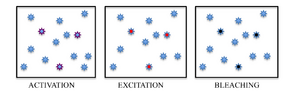
Immobilized fluorescent proteins being photoactivated, excited and bleached
-
author= Izeddin I, Specht CG, Lelek M, Darzacq X, Triller A, et al.
-
Investigating Sub Spine Actin Dynamics in Rat Hippocampal Neurons with Super Resolution Optical Microscopy [9]
المصادر
- ^ E. Betzig, G. H. Patterson, R. Sougrat, O. W. Lindwasser, S. Olenych, J. S. Bonifacino, M. W. Davidson, J. Lippincott-Schwartz, H. F. Hess (2006). "Imaging Intracellular Fluorescent Proteins at Nanometer Resolution". Science. 313 (5793): 1642–1645. Bibcode:2006Sci...313.1642B. doi:10.1126/science.1127344. PMID 16902090.
{{cite journal}}: CS1 maint: multiple names: authors list (link) - ^ S. T. Hess, T. P. Giriajan, M. D. Mason (2006). "Ultra-high resolution imaging by Fluorescence Photoactivation Localization Microscopy". Biophysical Journal. 91 (11): 4258–4272. Bibcode:2006BpJ....91.4258H. doi:10.1529/biophysj.106.091116. PMC 1635685. PMID 16980368.
{{cite journal}}: CS1 maint: multiple names: authors list (link) - ^ M. J. Rust, M. Bates, X. Zhuang (2006). "Sub diffraction-limit imaging by stochastic optical reconstruction microscopy (STORM)". Nature Methods. 3 (20): 793–796. doi:10.1038/nmeth929.
{{cite journal}}: CS1 maint: multiple names: authors list (link) - ^ "Method of the Year 2008". Nature Methods. 6 (1): 1–109. 2009. doi:10.1038/nmeth.f.244.
- ^ Ha, Taekjip and Tinnefeld, Philip (2012). "Photophysics of Fluorescent Probes for Single-Molecule Biophysics and Super-Resolution Imaging". Annual Review of Physical Chemistry. 63 (1): 595–617. Bibcode:2012ARPC...63..595H. doi:10.1146/annurev-physchem-032210-103340.
{{cite journal}}: CS1 maint: multiple names: authors list (link) - ^ Bo Huang and Hazen Babcock and Xiaowei Zhuang (2010). "Breaking the Diffraction Barrier: Super-Resolution Imaging of Cells". Cell. 143 (7): 1047–58. doi:10.1016/j.cell.2010.12.002.
- ^ Shtengel, Gleb and Galbraith, James A. and Galbraith, Catherine G. and Lippincott-Schwartz, Jennifer and Gillette, Jennifer M. and Manley, Suliana and Sougrat, Rachid and Waterman, Clare M. and Kanchanawong, Pakorn and Davidson, Michael W. and Fetter, Richard D. and Hess, Harald F. (2009). "Interferometric fluorescent super-resolution microscopy resolves 3D cellular ultrastructure". Proceedings of the National Academy of Sciences. 106 (9): 3125–3130. Bibcode:2009PNAS..106.3125S. doi:10.1073/pnas.0813131106. PMC 2637278. PMID 19202073.
{{cite journal}}: CS1 maint: multiple names: authors list (link) - ^ Greenfield D, McEvoy AL, Shroff H, Crooks GE, Wingreen NS; et al. (2009). "Self-Organization of the Escherichia coli Chemotaxis Network Imaged with Super-Resolution Light Microscopy". PLoS Biology. 7 (6). doi:10.1371/journal.pbio.1000137.
{{cite journal}}: Explicit use of et al. in:|author=(help)CS1 maint: multiple names: authors list (link) CS1 maint: unflagged free DOI (link) - ^ Tatavarty V, Kim E, Rodionov V, Yu J (2009). "Investigating Sub-Spine Actin Dynamics in Rat Hippocampal Neurons with Super-Resolution Optical Imaging". PLoS ONE. 4 (11): e7724. Bibcode:2011PLoSO...615611I. doi:10.1371/journal.pone.0015611.
{{cite journal}}: CS1 maint: multiple names: authors list (link) CS1 maint: unflagged free DOI (link)
وصلات خارجية
- Superresolution Microscopy within Zeiss educational page in Microscopy and Digital Imaging
- Fundamental Concepts in Super Resolution within Nikon educational resources for Microscopy Education
- Eric Betzig and Harald Hess talk: Developing PALM Microscopy
- Xiaowei Zhuang talk: Super-Resolution Microscopy
المجهرية الضوئية | ||
|---|---|---|
| Illumination and contrast methods |  | |
| وسائل الاستشعاع | ||
| Sub-diffraction limit techniques | ||