الحاجز بين البطينين
| Interventricular septum | |
|---|---|
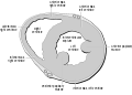
 Section of the heart showing the ventricular septum. | |
 Interior dorsal half of heart of nearly 5 weeks old human embryo. (Labeled as 'septum inferius') | |
| Details | |
| جزء من | Heart |
| الشريان | anterior interventricular branch of left coronary artery and Posterior interventricular artery |
| Identifiers | |
| اللاتينية | septum interventriculare cordis |
| MeSH | D054088 |
| TA98 | A12.1.00.013 |
| TA2 | 3970 |
| FMA | 7133 |
| المصطلحات التشريحية | |
الحاجز بين البطينين أو الحاجز البطيني (interventricular septum) هو عبارة عن جدار قوي يفصل بين بطيني القلب. يتجه الحاجز بين البطينين بشكل مائل نحو الخلف والأيمن وتتوافق حوافه مع الثلمين بين البطينين الأمامي والخلفي.
The ventricular septum is directed obliquely backward to the right and curved with the convexity toward the right ventricle; its margins correspond with the anterior and posterior interventricular sulci. The lower part of the septum, which is the major part, is thick and muscular, and its much smaller upper part is thin and membraneous.[1]
During each cardiac cycle the interventricular septum contracts by shortening longitudinally and becoming thicker.
البنية

The interventricular septum is the stout wall separating the ventricles, the lower chambers of the heart, from one another.
The ventricular septum is directed obliquely backward to the right and curved with the convexity toward the right ventricle; its margins correspond with the anterior and posterior longitudinal sulci. The greater portion of it is thick and muscular and constitutes the muscular interventricular septum. Its upper and posterior part, which separates the aortic vestibule from the lower part of the right atrium and upper part of the right ventricle, is thin and fibrous, and is termed the membranous ventricular septum.
أقسامه
يتشكل الحاجز بين البطينين من قسم:
- القسم العضلي وهو الأكبر
- القسم الغشائي وهو قسم صغير يشكل الجزء العلوي الخلفي
Blood supply
The posterior interventricular artery, a branch of right coronary artery, supplies the posterior 1/3 of the interventricular septum. The remaining anterior 2/3 is supplied by the anterior interventricular artery, which is a septal branch of the left anterior descending artery, which is a branch of left coronary artery. [2]
Development
The muscular part of the interventricular septum derives from the bulboventricular flange which is developed due to differential growth of primitive ventricle and bulbous cordis. Membranous part has a neural crest origin which connects the upper free margin of the bulboventricular flange and anterior and posterior endocardial cushions of atrio ventricular canal. It also gets attached to lower border of spiral septum or the aorticopulmonary septum.
In the final stages of the heart development, the interatrial septum aligns in the same plane as the interventricular septum. The gap between the interatrial septum and interventricular septum forms the membranous part of interventricular septum. [3]
الأهمية السريرية
A ventricular septal defect (VSD), a hole in the interventricular septum is one of the four congenital defects of the condition of tetralogy of Fallot. A VSD can cause a left-to-right shunt of blood flow in the heart and is one of the most common of the congenital heart defects. This type of shunt is an acyanotic disorder that can result in ventricular hypertrophy. [4]
The alignment of interventricular septum and interatrial septum is disturbed in various congenital heart diseases.[5]
عند وجود فتحة في الحاجز بين البطينين يدعى ذلك عيب الحاجز البطيني.
صور إضافية
Heart normal short axis echo

انظر أيضاً
المراجع
- ^ "Interventricular septum".
- ^ Futami, C.; Tanuma, K.; Tanuma, Y.; Saito, T. (1 April 2003). "The arterial blood supply of the conducting system in normal human hearts". Surgical and Radiologic Anatomy (in الإنجليزية). 25 (1): 42–49. doi:10.1007/s00276-002-0085-7. ISSN 1279-8517. PMID 12819949. S2CID 24687834. Retrieved 13 December 2022.
- ^ MORRIS EW (December 1957). "The interventricular septum". Thorax. 12 (4): 304–12. doi:10.1136/thx.12.4.304. PMC 1019182. PMID 13496033.
- ^ Pillitteri, Adele (2010). Maternal & Child Health Nursing: Care of the Childbearing & Childrearing Family (in الإنجليزية). Lippincott Williams & Wilkins. ISBN 9781582559995.
- ^ Yoo, Shi‐Joon; Saito, Mika; Hussein, Nabil; Golding, Fraser; Goo, Hyun Woo; Lee, Whal; Lam, Christopher Z.; Seed, Mike; Dragulescu, Andreea (2020-11-17). "Systematic Approach to Malalignment Type Ventricular Septal Defects". Journal of the American Heart Association (in الإنجليزية). 9 (22): e018275. doi:10.1161/JAHA.120.018275. ISSN 2047-9980. PMC 7763733. PMID 33170057.
وصلات خارجية
- علم الأنسجة في جامعة اوكلاهوما 128_06 - "Heart and semilunar valve"